
Did you know? The global market of cancer immunotherapy is projected to be worth over $296.01 billion in 2033. But where each approved cancer drug costs an average of $648 million to develop, this treatment only works for 20% to 40% of patients.
Also, late-stage clinical failures drain resources. Unpredictable responses frustrate investors and patients alike. The answer to this challenge is immune profiling in cancer.
Immune Profiling in cancer decodes how tumors interact with the immune system. They analyze complex immune signatures to predict treatment outcomes. The results are impressive: AI-driven models now predict treatment responses with 70-80% accuracy.
In this article, we will explore techniques for immune profiling in cancer research and how you can leverage these technologies to revolutionize drug development.
Key Takeaways
- Immune profiling in cancer helps predict treatment responses by analyzing how tumors interact with the immune system. AI models now predict immunotherapy outcomes with 70-80% accuracy.
- Key techniques include flow cytometry/CyTOF for detailed cellular analysis, single-cell RNA sequencing for detecting tumor diversity, spatial profiling for tissue context, liquid biopsy for non-invasive monitoring, and proteogenomics for in-depth molecular data.
- Recent breakthroughs include personalized mRNA vaccines cutting melanoma recurrence by 44%, AI biomarkers outperforming FDA tests, and the first FDA-approved TIL therapy for solid tumors.
- Technical challenges still exist, including sample quality issues, data complexity, lack of standardization, and gaps in validation that slow clinical application and raise research costs.
- Biostate AI provides a solution starting at $80/sample with AI-powered tools that remove technical barriers, allowing researchers to focus on discovery, not data processing.
What is Immune Profiling?
Immune profiling identifies and measures immune cell populations based on their characteristics and activity.
At its core, immune profiling in cancer reveals which immune cells are present, how many exist, what roles they play, and how they communicate. By examining tissues, blood, or the tumor microenvironment, researchers gain a clearer view of the immune landscape.
Components of Immune Profiling
In immune profiling, we analyze various components and aspects of the immune system at different levels, such as:
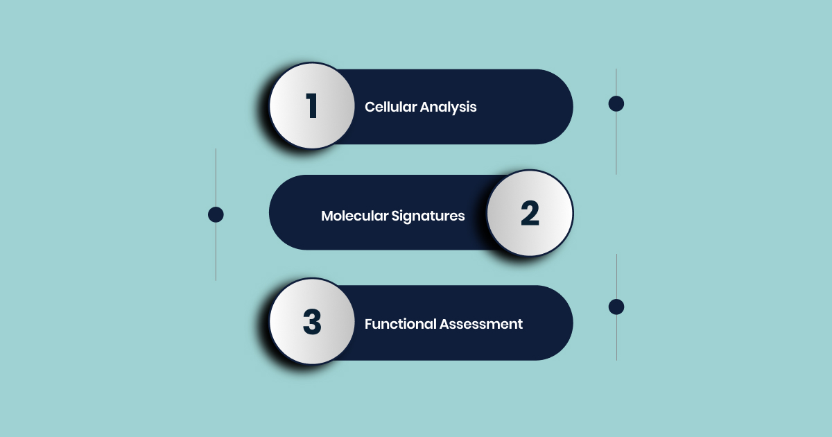
1. Cellular Analysis
Immune profiling identifies different types of immune cells, including T cells, B cells, natural killer (NK) cells, macrophages, neutrophils, and dendritic cells. Each cell type plays distinct roles in immune responses. The analysis measures both their quantity and activation state.
2. Molecular Signatures
Beyond counting cells, immune profiling examines the molecules these cells produce. This includes cytokines (immune signaling proteins), chemokines (cell recruitment signals), and surface markers that indicate cell function. These molecular signatures reveal whether the immune system is activated, suppressed, or in a balanced state.
3. Functional Assessment
Modern immune profiling goes beyond static snapshots. It measures how immune cells respond to stimuli, their metabolic activity, and their ability to communicate with other cells.
It distinguishes “hot” tumors rich in immune cells from “cold” tumors with little infiltration, guiding personalized treatment strategies. This functional dimension is crucial for understanding immune system effectiveness in cancer research.
For pharmaceutical research, immune profiling supports patient selection in clinical trials, identifies biomarkers, and explains treatment mechanisms. AI-driven models like Prognosis AIcan predict responses with 70–80% accuracy, turning uncertain immune reactions into actionable insights.
With this foundational understanding of immune profiling and its core components, let’s explore the specific techniques that make these detailed analyzes possible in cancer research.
Techniques for Immune Profiling in Cancer
Immune profiling in cancer research involves analyzing the immune cells present within the tumor microenvironment and peripheral blood to understand their type, quantity, activation status, and interactions with cancer cells.
Here’s an overview of the key techniques used in immune profiling:
Flow Cytometry and Mass Cytometry (CyTOF): High-Dimensional Cellular Analysis
Flow cytometry uses lasers and fluorescent antibodies to measure cell size, structure, and protein expression. Newer spectral flow cytometry detects 40+ antigens at once.
Mass cytometry (CyTOF) replaces fluorescent tags with metal isotopes, allowing 40+ markers to be profiled simultaneously with minimal noise. Millions of cells can be analyzed daily, making CyTOF especially valuable for immune mapping and biomarker discovery.
Why It Matters for Pharma
These tools provide high-throughput, protein-level data to:
- Identify immune cell subsets driving tumor response or suppression
- Run functional assays for therapies like ADCs and CAR-T
- Support cost-efficient deep profiling (~$0.003 per cell in CyTOF)
Drug Development Applications
Flow cytometry is central to CAR-T therapies, from manufacturing quality checks and functional assays to safety monitoring and post-infusion tracking.
For instance,
- Carvykti’s clinical development relied heavily on flow cytometry to monitor CAR-T expansion and patient response.
- CyTOF, meanwhile, enables broad immune profiling across T cells, B cells, NK cells, and more, helping refine biomarker strategies and receptor occupancy assays.
Challenges
- Solid tumors must be broken into single-cell suspensions, which is time-intensive and requires expertise.
- Mass cytometry depends on available metal-tagged antibodies, which limits flexibility.
- Large datasets can be challenging to analyze, though newer workflows are reducing this burden.
Using the same platforms across preclinical and clinical stages strengthens translation, speeds biomarker validation, and reduces trial risks. Beyond research, flow cytometry is a manufacturing must-have, ensuring safety, potency, and compliance for cell therapies. For pharma, this makes it both a discovery engine and an operational backbone.
While flow cytometry and CyTOF excel at analyzing known cell populations at scale, understanding tumor complexity often requires looking deeper at individual cells and their unique molecular signatures.
Single-Cell Immune Profiling: Dissecting Tumor Heterogeneity and Rare Cell Populations
Traditional bulk RNA-seq averages gene expression across cells, hiding important cell-type differences and tumor heterogeneity. Whereas, single-cell RNA sequencing (scRNA-seq) provides a clearer picture, showing tumor complexity at single-cell resolution.
Key Capabilities
- Unbiased discovery: Identifies new cellular subtypes and states by analyzing thousands of genes per cell.
- Rare cell detection: Captures uncommon but biologically important populations.
- TCR integration: Technologies like 10x Genomics’ Chromium allow scRNA-seq and TCR-seq together, enabling analysis of clonal expansion and immune responses in the tumor microenvironment.
- Spatial integration: While single-cell dissociation removes spatial context, combining with spatial transcriptomics restores this layer of insight.
Applications in Drug Development
- Biomarker discovery: scRNA-seq identified GZMK-expressing CD8+ T cells as predictors of checkpoint blockade response in AML.
- Drug resistance insights: Tracks transcriptional changes in response to therapy, highlighting resistant populations and mechanisms.
- New drug targets: Identifies cancer cell states driving resistance or progression.
- Cell therapy optimization: Helps improve TIL therapy by showing how oncolytic viruses can make tumor cells act like antigen-presenting cells, boosting T-cell activation.
- snRNA-seq advantage: Works on frozen samples, enabling the use of archived clinical trial tissues for new analysis and collaboration.
Strengths
The greatest strength of single-cell profiling is its ability to reveal rare or subtle cell states that bulk methods miss.
- These hidden populations often explain why some patients respond to therapy while others develop resistance.
- For pharma and biotech, this increases the chance of finding specific, actionable biomarkers and reduces failures from “one-size-fits-all” therapies.
- Frozen biopsy samples from past clinical trials hold untapped value. snRNA-seq makes it possible to analyze these archives, generating insights into drug response and resistance without needing fresh samples.
This approach helps validate biomarkers, uncover new indications, and lower R&D costs while accelerating discovery.
Limitations
- Requires tissue dissociation, losing spatial structure.
- Needs specialized equipment, expertise, and rigorous data processing.
- Dependent on sample quality and sequencing depth.
Although single-cell analysis reveals remarkable cellular diversity, it comes with a trade-off: the loss of spatial context that tells us where these cells interact within the tumor microenvironment.
Spatial Immune Profiling: Contextualizing Immune Interactions in the TME
Understanding where immune cells sit in tissue, which cells they interact with, and what genes or proteins they express is key to decoding immune behavior. The success of immunotherapy often depends not only on which immune cells are present but also on their spatial organization inside the tumor microenvironment (TME).
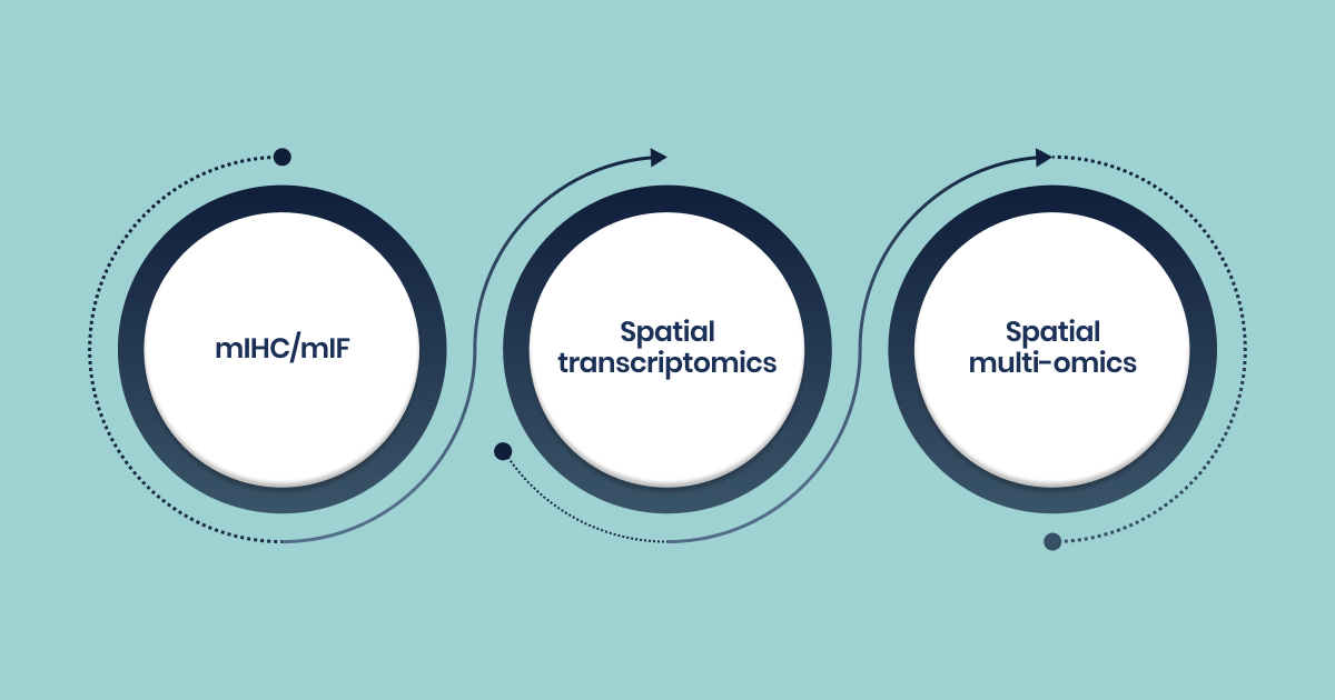
Key Technologies
- mIHC/mIF: Multiplex immunohistochemistry and immunofluorescence stain multiple markers at once, defining complex immunophenotypes and quantifying immune cells in tissue.
- Spatial transcriptomics: Maps gene expression within intact tissue sections, preserving natural structure. Examples: 10x Genomics Visium, Vizgen’s MERSCOPE Ultra.
- Spatial multi-omics: Integrates RNA, DNA, and protein data with spatial context, providing a 3D view of tumor heterogeneity.
Applications in Drug Development
- TME characterization: Defines immune escape mechanisms and predicts therapeutic response.
- Predictive biomarkers: mIHC/mIF outperforms PD-L1 IHC or TMB in predicting checkpoint inhibitor response. For example, intratumoral CD8+CD39+ density or CD8+FoxP3+ ratios in NSCLC correlate with outcomes.
- Therapy guidance: Distinct HCC spatial immunotypes (depleted, compartmentalised, enriched) show different progression-free survival under ICI. Each requires tailored strategies, such as co-targeting exhaustion markers, stroma-directed therapy, or local immune priming.
- Overcoming barriers: Spatial data shows how fibroblasts (αSMA+) can physically block CD8+ T-cell infiltration in NSCLC, explaining poor immunotherapy response. This suggests stroma-targeting drugs could improve infiltration.
- Resistance monitoring: Spatial transcriptomics applied to banked samples helps explain relapse or resistance, providing retrospective insights.
Strengths
Single-cell methods show immune cell types and molecular states. Spatial methods add the missing layer, showing how cells are organized and interacting.
- In hepatocellular carcinoma (HCC), CD8 T-cell numbers alone are not predictive. Their location, tumor core vs stroma, creates distinct immunotypes with different therapy outcomes. For pharma and biotech, this means spatial biomarkers should be part of clinical trial design and companion diagnostics.
- Spatial profiling shows that immune suppression is not only molecular but also architectural.
- Fibroblast walls or extracellular matrix can isolate T cells from tumor cells, blocking immune attack.
- This opens new drug discovery paths: therapies that dismantle stromal barriers could combine with checkpoint inhibitors to improve efficacy.
Limitations
- Complex datasets require advanced computational analysis.
- Lack of standardization in staining and imaging protocols.
- Ongoing harmonization efforts are addressing reproducibility across labs.
While tissue-based spatial profiling provides rich contextual information, accessing tumor tissue repeatedly for monitoring treatment response presents practical challenges that have driven innovation in blood-based approaches.
Liquid Biopsy: Non-Invasive Monitoring and Dynamic Biomarker Detection
Liquid biopsy is a minimally invasive method that detects tumor-derived material in blood, such as ctDNA, cfDNA, RNA, and circulating tumor cells (CTCs). It is especially useful when tissue samples are unavailable or hard to obtain.
Advantages for Pharma and Biotech
- Enables serial, real-time monitoring of tumor burden and genetic changes.
- Provides comprehensive genomic profiling from a single blood draw (driver mutations, resistance markers, SNVs, indels, fusions).
- Detects resistance or recurrence earlier than imaging, giving lead time for treatment adjustment.
- Captures tumor heterogeneity missed by single-site biopsies.
Applications in Drug Development
- Response monitoring: Longitudinal ctDNA assessment correlates with ICI outcomes (OS, PFS, durability).
- Therapy guidance: Identifies driver oncogenes and acquired resistance mutations to inform next-line therapy.
- Stratification: Differentiates patients suitable for curative vs palliative approaches.
- MRD detection: Identifies residual disease not visible by imaging. FDA-approved liquid biopsy assays already support precision oncology.
- Clinical trials: ctDNA-adaptive designs enable dynamic interventions, and the FDA has issued draft guidance on ctDNA use in solid tumors.
Strengths
- Liquid biopsy allows frequent molecular readouts, enabling adaptive trials guided by ctDNA clearance or resistance emergence.
- This can shorten trial timelines, lower costs, and reduce patient drop-out.
- It also supports “living drugs” through continuous monitoring.
- For patients unable to undergo tissue biopsy, liquid biopsy provides a practical alternative, expanding access to molecular profiling and targeted therapies.
- For companies, it broadens trial eligibility and market reach while aligning with efforts to improve equity and access in oncology.
Limitations
- Sensitivity challenges in early-stage disease (low ctDNA levels) and difficulty detecting fusions/CNAs.
- False positives may arise from clonal haematopoiesis.
- Negative results require cautious interpretation.
As powerful as liquid biopsy is for monitoring tumor dynamics, it primarily captures genetic information. To fully understand how these genetic changes translate into functional differences that affect treatment response, we need approaches that bridge the gap between genes and proteins.
Proteogenomics: Bridging Genotype and Phenotype
Proteogenomics integrates genomics (gene-level data) and proteomics (protein-level data) to provide a holistic view of tumors. While genomics and transcriptomics reveal what genes are present or expressed, proteomics measures functional proteins, their abundance, modifications, and interactions, which directly drive cellular behavior and therapy response.
This makes proteogenomics a key bridge between genotype and phenotype, offering deeper insights into tumor biology.
Key Capabilities
- Molecular Profiling: Assesses genetic alterations, protein abundance, interactions, and modifications.
- Multi-Omics Integration: Combines genomic, transcriptomic, and proteomic data for a systems-level view.
- tumor Subtyping: categorizes tumors (immune hot, warm, cold) based on immune and stromal composition.
Applications in Cancer Drug Development
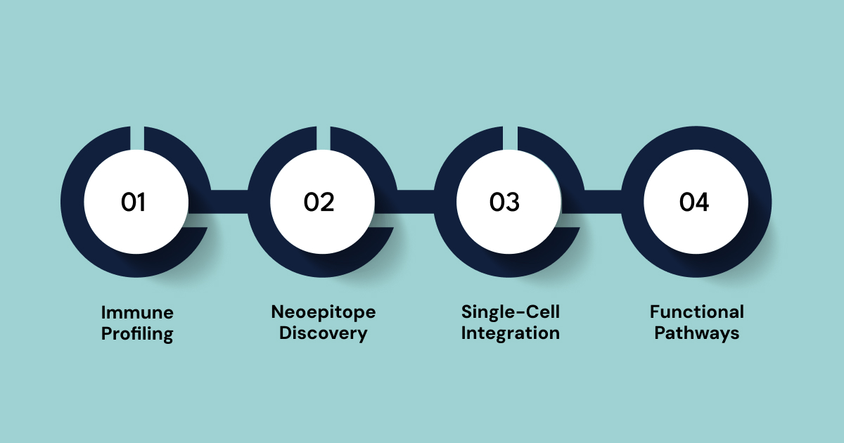
- Immune Profiling: Proteogenomics deconstructs TME composition, revealing immune densities that guide therapy. Example: PDAC proteogenomics identified “immune-cold” tumors with glycolysis upregulation and reduced endothelial adhesion, suggesting metabolism and vascular targeting as strategies to boost immunity.
- Neoepitope Discovery: Mass spectrometry identifies tumor-specific antigens from mutations (e.g., FGFR2 fusions in iCCA) for immunotherapy development.
- Single-Cell Integration: Enhances bulk data with finer resolution. In TNBC, CXCL13+ T cell clusters identified via scRNA-seq predicted response to PD-L1 blockade.
- Functional Pathways: Goes beyond gene expression to uncover dysregulated protein-driven pathways (e.g., VEGF, glycolysis), highlighting new drug targets.
Pharma/Biotech Value
- Precision Targeting: Enables drug discovery against functional protein-level changes driving resistance.
- Expanding ICI Use: characterizes “immune-cold” tumors and identifies strategies to make them “hot,” opening opportunities for combination therapies.
- Translational Impact: Findings (biomarkers, targets) validated in vitro/in vivo can be incorporated into clinical assays and companion diagnostics.
Proteogenomics provides a functionally relevant layer of cancer biology that genomics alone cannot capture. For drug developers, it offers a roadmap to identify new targets, improve biomarker precision, and design therapies tailored to the immune context, especially for tumors resistant to current immunotherapies.
These established techniques have laid the groundwork for remarkable advances in the field. The convergence of improved methodologies, artificial intelligence, and clinical validation has produced breakthrough discoveries that are reshaping cancer treatment.
Recent Breakthroughs and Emerging Trends in Immuno-Oncology
Immuno-oncology has changed cancer treatment by using the body’s immune system to attack tumors. Between 2023 and 2025, major breakthroughs are shaping how these therapies are developed and used. These advances are making treatments more precise, more effective, and applicable to a wider range of cancers.
| Breakthrough Area | Specific Example | Key Impact/Result | Implications for Immune Profiling |
| RNA Cancer Vaccines | mRNA-4157 (V940) + Pembrolizumab in Melanoma | 44% reduction in recurrence riskSuperior 3-year recurrence-free survival | Drives the need for AI-driven neoantigen selectionPatient-specific immune profiling and real-time immune monitoring |
| Personalized mRNA vaccine for Pancreatic Cancer | Remarkable efficacyVaccine-induced immune responses persisting for nearly 4 years | Validates personalized vaccine approach for historically resistant cancersEmphasizes deep immune response analysis | |
| Glioblastoma mRNA vaccine (UF-developed) | Reprogrammed the immune system to attack the tumor within 48 hoursConverted “cold” to “hot” tumors | Highlights the role of profiling in understanding TME modulation and immune cell infiltration | |
| AI-Driven Biomarkers | SCORPIO AI tool | More accurate than FDA-approved tests (PD-L1, TMB) in predicting tumor shrinkage and survival | Emphasizes multi-parametric data integration (clinical, imaging, multi-omics) for robust predictive biomarkers |
| Multi-LLM AI Frameworks for Biomarker Discovery | Enhances robustness, reproducibility, and interpretability of gene selectionMinimizes single-model bias | Accelerates target identification and validationRequires expertise in complex AI/ML platforms | |
| AI platforms for precision immune cell design | Reduces drug development time from years to weeks for targeted immune cell therapies | Transforms early-stage drug discoveryEnables rapid iteration of therapeutic candidates | |
| Advanced Cell Therapies | First FDA-approved TIL therapy (lifileucel) for advanced melanoma | Fundamental shift towards personalized interventions for solid tumors | Requires robust immune profiling for patient selectionProduct characterization and monitoring |
| First TCR-engineered therapy (afamitresgene autoleucel) for solid tumor | Expands cell therapy applicability to solid tumorsHighlights the importance of TCR engineering | Necessitates high-resolution HLA typing and TCR repertoire analysis for patient eligibility and safety | |
| CAR-T Therapy (Carvykti) monitoring | Flow cytometry tracked CAR-T cell expansion and efficacy in multiple myeloma | Demonstrates the critical role of flow cytometry in manufacturing QCFunctional assessment and post-infusion monitoring |
Despite these exciting advances, the path from laboratory innovation to clinical implementation remains complex, with several persistent challenges that researchers must navigate.
Technical Challenges in Immune Profiling in Cancer
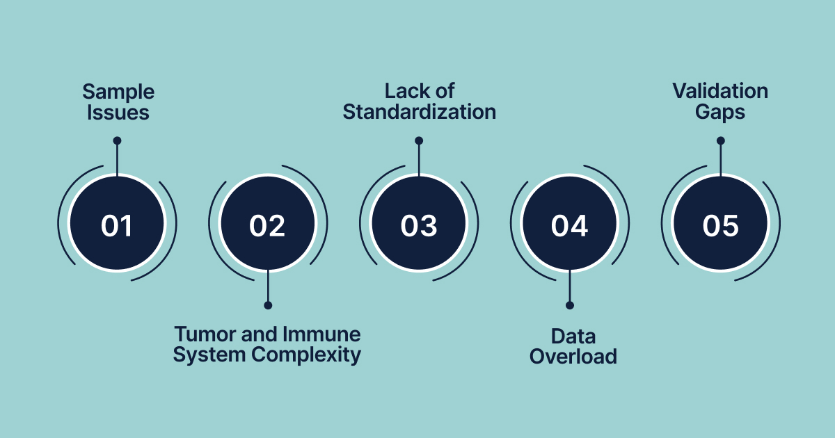
Immune profiling in cancer comes with several technical challenges that can slow progress and affect results:
- Sample Issues: Getting high-quality tissue samples is tough. Archival or fixed samples (like FFPE) are often less reliable than fresh ones, and repeated sampling during treatment is rarely possible. Many patients cannot provide enough tissue for multiple tests.
- Tumor and Immune System Complexity: Tumors and their surrounding immune cells are highly complex and vary from patient to patient. This heterogeneity makes it hard to identify clear biomarkers or predict responses.
- Lack of Standardization: There are no universal standards for how immune profiling is done or how results are interpreted. Different labs may get different results from the same sample.
- Data Overload: New techniques like single-cell sequencing and spatial profiling generate massive amounts of complex data. Making sense of it—which patterns matter and why—is a significant challenge and requires advanced, costly analysis tools.
- Validation Gaps: Few immune profiling markers have been fully validated for routine clinical use. Most are still experimental and not ready for everyday decision-making.
These challenges highlight the need for integrated solutions that can streamline the immune profiling workflow while maintaining scientific rigor and reducing barriers to discovery.
How Biostate AI Can Simplify Your Cancer Research?
Immune Profiling in cancer faces significant obstacles that can derail even the most promising research initiatives. Many researchers spend more time dealing with technical hurdles than focusing on scientific discovery.
Biostate AI changes this by offering an integrated platform that handles the entire research process, from sample collection to actionable insights. Our solution streamlines cancer research workflows, combining high-quality RNA sequencing with AI-powered tools tailored for immune profiling.
Key Features That Accelerate Your Cancer Research:
- Cost-Effective Sequencing: High-quality RNA sequencing from just $80/sample with results in 1–3 weeks.
- Flexible Sample Compatibility: Works with various sample types—blood, tissue, culture, or purified RNA, requiring as little as 10µL of blood or 10 ng of RNA.
- Low-Quality Sample Processing: Can handle degraded samples with RIN as low as 2, ideal for archived cancer specimens.
- Comprehensive Transcriptome Coverage: Full RNA-seq analysis covering both mRNA and non-coding RNA to map the immune landscape fully.
- AI-Powered Analysis: OmicsWeb offers easy, no-code data analysis with an AI assistant to interpret complex results.
- Multi-Omics Integration: Combine RNA-seq, WGS, methylation, and single-cell data for a deeper immune profile.
- Predictive Disease Modeling: Biobase AI predicts drug toxicity with 89% accuracy and therapy selection with 70% accuracy, turning clinical samples into actionable models.
- Automated Workflows: Streamlined pipelines deliver publication-ready insights, eliminating manual bottlenecks.
Biostate AI brings all these capabilities into one platform, allowing researchers to focus on scientific progress rather than technical challenges, accelerating the path from immune profiling data to therapeutic breakthroughs.
Final Words
Immune profiling in cancer is a key part of precision oncology, offering deep insights into how tumors interact with the immune system. With tools like flow cytometry, CyTOF, single-cell sequencing, and spatial profiling, researchers can now decode immune landscapes and predict how tumors will respond to treatments.
However, challenges like sample quality issues, data complexity, and lack of standardization still slow down progress and limit clinical applications.
Biostate AI removes both financial and technical barriers to breakthrough research, starting at just $80 per sample with quick results. We combine high-quality RNA sequencing with AI-powered analysis tools like OmicsWeb and Biobase predictive models, allowing researchers to turn complex immune data into actionable insights with no bioinformatics expertise or heavy infrastructure required.
Ready to speed up your cancer immune profiling research while cutting costs and complexity? Get your quote today and see how Biostate AI can streamline your research pipeline from sample to insight.
FAQs
Q: How do I ensure regulatory compliance when implementing immune profiling in clinical trials?
A: Follow Good Laboratory Practices (GLP) for FDA submissions and validate your assays according to bioanalytical guidelines. Document standard operating procedures (SOPs) for sample collection, processing, and analysis. For companion diagnostics, engage with the FDA early through pre-submission meetings. European regulations (EMA) have similar requirements but may differ in technical details. Consider consulting with regulatory experts for guidance on approval processes.
Q: What are the key considerations for biomarker validation and transitioning to clinical-grade assays?
A: Biomarker validation involves analytical validation (precision, accuracy), clinical validation (outcome correlation), and clinical utility (impact on patient care). To transition to clinical-grade assays, ensure analytical performance (detection limits, reproducibility) and demonstrate robustness in patient samples. Show that the assay is actionable for clinical decisions and plan for smooth transfer to clinical labs with quality control measures in place.
Q: How can I design studies to demonstrate clinical utility to payers and regulators?
A: Design studies that link biomarker results to outcomes like survival, progression-free survival, or quality of life. Include cost-effectiveness analysis to show how immune profiling reduces unnecessary treatments or optimizes resources. Use appropriate statistical power for key endpoints and consider adaptive trial designs to demonstrate utility efficiently. Engage payers early to understand their requirements for evidence supporting coverage decisions.
Q: What intellectual property considerations should I keep in mind when developing immune profiling technologies or biomarkers?
A: Perform a patent landscape analysis to avoid infringement and identify innovation opportunities. Consider intellectual property for gene signatures, algorithms, data analysis methods, and diagnostics. File provisional patents early, especially for new biomarker combinations or AI-driven approaches. Be mindful of existing patents and licensing agreements, and work with IP attorneys to protect your innovations and navigate patent filings for international markets.