
Researchers often face challenges in capturing cellular diversity, especially in diseases and treatment resistance, as traditional bulk RNA sequencing falls short. Single-cell RNA sequencing (scRNA-seq) has revolutionized gene expression studies by analyzing individual cells.
However, it comes with hurdles like the need for immediate tissue processing, logistical issues, and harsh dissociation protocols that can damage delicate cells. These challenges are especially pronounced with tough tissues like the brain, kidney, or fibrotic samples, where enzymatic digestion can distort data.
This is where single-nucleus RNA sequencing (snRNA-seq) offers a breakthrough. It’s more flexible, allowing researchers to work with frozen or archived tissues, removing the pressure for immediate processing. This makes snRNA-seq ideal for retrospective studies.
In this article, we’ll compare scRNA-seq and snRNA-seq, highlighting how snRNA-seq enhances rare cell detection and improves data quality.
TL;DR
- scRNA-seq offers high-resolution insights into gene expression from individual cells, while snRNA-seq, focusing on nuclei, excels in handling difficult tissues and archived samples.
- snRNA-seq improves rare cell detection and reduces dissociation bias, especially in fragile tissues like neurons, compared to scRNA-seq’s higher sensitivity for immune cells.
- scRNA-seq is ideal for fresh tissues, while snRNA-seq offers more flexibility, handling frozen or archived samples with greater accuracy in challenging cell types like neurons.
What is Single-Cell RNA-seq and Single-Nuclei RNA-seq
Single-cell and single-nucleus RNA sequencing (scRNA-seq and snRNA-seq) have revolutionized molecular biology, enabling detailed analysis of cellular heterogeneity and dynamics. Unlike traditional bulk RNA sequencing, which provides an average expression profile across many cells, these technologies offer insights into individual cell or nucleus gene expression, uncovering complex cellular states, transitions, and subpopulations.
What is scRNA-seq
Single-cell RNA sequencing (scRNA-seq) provides a high-resolution view of gene expression at the single-cell level. It involves isolating individual cells, extracting and amplifying mRNA, and performing Next-Generation Sequencing (NGS).
By replacing population-averaged data with single-cell resolution, scRNA-seq provides unparalleled insight into gene expression dynamics and disease mechanisms.
What is snRNA-seq
Single-nucleus RNA sequencing (snRNA-seq), also referred to as single nuclei RNA sequencing or sNuc-seq, offers an alternative RNA sequencing methodology that profiles gene expression from isolated cell nuclei rather than intact whole cells.
Advantages
- This is a critical method when whole-cell dissociation is challenging or damaging.
- This method is particularly useful for tissues that are hard to dissociate, such as neurons or adipocytes, or archived frozen samples.
- One key advantage of snRNA-seq is its ability to minimize spurious gene expression, as mature ribosomes in the cytoplasm are not available to translate mRNAs post-dissociation, ensuring more accurate transcriptional data.
By focusing on the nucleus it provides a robust and less biased method to access transcriptomic information from contexts previously inaccessible to single-cell resolution studies.
While both methods revolutionize our understanding of gene expression, the practical applications of each can differ significantly depending on the sample type. Let’s explore how these two technologies measure up when it comes to rare cell detection and data quality.
How Efficient is snRNA-seq versus scRNA-seq for Rare Cell Detection and Data Quality
scRNA-seq, due to its reliance on whole-cell dissociation, often captures reactive and immune cells more efficiently but struggles with certain fragile or difficult-to-dissociate populations like specific neurons.
In contrast, snRNA-seq, with its less disruptive nuclear isolation process, provides more balanced coverage of certain cell types, especially neurons, but may miss immune cells.
Here is the key comparison between these two sequencing technologies:
| Feature | scRNA-seq Performance | snRNA-seq Performance |
| Rare Cell Detection Efficiency | High resolution, but can miss fragile/embedded rare cells due to dissociation bias and cell loss | Improved capture of difficult-to-isolate and rare cell types (e.g., neurons, adipocytes, fibrotic glia) due to nuclear robustness |
| Dissociation Bias/Cell Loss | Significant bias and cell loss, especially for cells strongly embedded or fragile; underrepresentation of certain cell types | Less biased cellular coverage; nuclei are more resistant to dissociation, reducing cell loss |
| Compatibility with Challenging Samples | Primarily requires fresh, viable tissue; not suitable for frozen/fixed samples | Highly compatible with fresh, frozen, or fixed tissues, including archived biobank samples |
| RNA Content Captured | Captures cytoplasmic and nuclear transcripts (total mRNA), leading to higher total UMIs and genes detected | Primarily nuclear transcripts (enriched for unspliced pre-mRNA and intronic sequences); lower total UMIs/genes but sufficient for cell type discrimination |
| Gene Detection Sensitivity | Generally higher (e.g., ~11,000 genes) | Generally lower (e.g., ~7,000 genes), but comparable for cell type discrimination when intronic reads are included |
| Mitochondrial/Ribosomal RNA Contamination | Higher levels can indicate cell damage or biological variation | Lower levels; presence serves as an excellent QC metric for nuclear stripping efficacy |
| Stress Response Artifacts | More susceptible to dissociation-induced transcriptional stress responses (e.g., IEGs) | Minimizes artificial transcriptional stress responses due to quick, mild nuclear dissociation |
While scRNA-seq and snRNA-seq each have distinct advantages and limitations, choosing the right method also depends heavily on the type of tissue being studied. Let’s examine how these technologies perform across different types of samples, especially in terms of compatibility and reliability.
Comparative Analysis: scRNA-seq vs. snRNA-seq
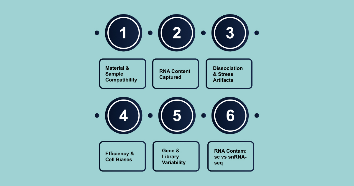
Here is a detailed comparison of scRNA-seq and snRNA-seq based on multiple criteria like sample compatibility, gene detection sensitivity, and biases in data collection. By categorizing these features, it will help you make an informed decision based on your experimental goals.
- Input Material and Sample Compatibility
Dissociation protocols, with distinct impacts on data fidelity, heavily influence the quality of transcriptomic profiles in scRNA-seq and snRNA-seq.
scRNA-seq requires fresh, intact cells in suspension, making it effective for freshly dissociated tissues. It struggles with frozen or fixed samples, particularly those from fragile cell types like neurons.
snRNA-seq, on the other hand, accommodates fresh, frozen, and fixed tissue samples, offering much greater flexibility. It is ideal for archived biobanks, post-mortem tissues, and challenging cell types like neurons, adipocytes, and heart cells.
Here is a table for sample compatibility and tissue suitability for scRNA-seq vs. snRNA-seq:
| Criterion | scRNA-seq Suitability/Limitations | snRNA-seq Suitability/Advantages |
| Fresh Tissue | Primarily effective for freshly dissociated tissues or cells in suspension | Highly flexible; accommodates fresh tissue |
| Frozen/Fixed Tissue | Very difficult to isolate single intact cells from frozen clinical samples; often incompatible | Offers substantial flexibility; accommodates frozen or fixed tissue samples; critical for biobanks and archived samples |
| Fragile Cells (e.g., Neurons) | Neurons are extremely fragile; unbiased neuronal profiling is difficult without freshly harvested, viable, intact neurons | Highly effective for analyzing complex and challenging cell types, including neurons; useful for postmortem brain samples |
| Difficult-to-Dissociate Tissues (e.g., Brain, Heart, Adipose) | Can be incompatible or require extensive preparation; it is difficult to dissociate heart tissue without damage | Accommodates a broader range of tissue types; effective for kidney, heart, and brain tissue; successful for entire mammalian hearts |
| Multi-nucleated Cells | Not explicitly mentioned as an advantage; likely challenging due to whole-cell isolation | Can uniquely interrogate multi-nucleated cells like trophoblasts, osteoclasts, and skeletal myocytes |
In summary, snRNA-seq is the preferred method for challenging, archived, or fragile tissue samples, while scRNA-seq remains ideal for fresh, dissociable cells. The choice of method depends on the sample type and research goals, with snRNA-seq offering broader compatibility and higher data fidelity for complex tissues.
- RNA Content Captured
scRNA-seq captures both cytoplasmic and nuclear RNA, focusing on mature, spliced mRNA. It provides insights into steady-state gene expression and active cellular processes like protein synthesis and metabolism, making it ideal for studying cell identity and functional states.
snRNA-seq isolates nuclear RNA, primarily unspliced pre-mRNA and intronic sequences, offering a deeper view of transcriptional regulation, gene activation, and RNA processing events like splicing. However, it does not capture cytoplasmic RNA, limiting detection of highly abundant transcripts or those with low nuclear presence.
| Criterion | scRNA-seq Characteristics | snRNA-seq Characteristics |
| RNA Types Captured | Cytoplasmic and nuclear transcripts (entire cell) | Primarily nuclear transcripts |
| Primary Biological Insights | Comprehensive view of mature, functional transcriptome; steady-state gene expression; active protein synthesis | Insights into nascent transcription, gene regulation, RNA processing events (e.g., splicing), and dynamic transcriptional changes |
| Enriched Functional Gene Categories | Protein synthesis, oxidative phosphorylation, viral gene expression, synapse formation, RNA metabolism, and splicing | Histone modification, GTPase activity, cellular morphogenesis, dendrite development |
scRNA-seq reflects the cell’s steady-state function, focusing on mature cytoplasmic transcripts. snRNA-seq, focusing on nuclear RNA, reveals transcriptional dynamics, splicing, and early gene regulation. While scRNA-seq is ideal for understanding cellular functions, snRNA-seq is more suitable for studying transcriptional processes and gene regulation.
- Dissociation Method and Stress Artifacts
scRNA-seq requires enzymatic digestion and mechanical disruption to isolate whole cells, which can cause stress responses (e.g., immediate early genes) and RNA degradation. These artifacts can obscure true biological signals, particularly in fragile cells like neurons, and require complex computational methods to clean the data, introducing additional biases.
However, snRNA-seq, by isolating only the nucleus, uses a milder, quicker dissociation process that reduces stress and preserves RNA integrity. This minimizes stress-induced artifacts, offering a more accurate representation of the transcriptome, especially in sensitive or difficult-to-isolate cells.
| Criterion | scRNA-seq Characteristics | snRNA-seq Characteristics |
| Primary Target for Isolation | Whole cells | Nuclei only |
| Dissociation Protocol Characteristics | Often complex protocols with long incubation times; requires robust enzymatic/mechanical disruption | Milder, quicker nuclear dissociation protocol; gentle lysis and centrifugation |
| Incidence of Transcriptional Stress Response | Prone to inducing biases due to cellular stress (e.g., IEGs); higher incidence of dissociation-induced stress | Significantly lower incidence of dissociation-induced transcriptional stress response |
| Risk of RNA Degradation | Can cause cell damage and RNA degradation | Efficient at preserving integrity by minimizing cellular stress and degradation |
| Protocol Complexity | Cumbersome and difficult; requires careful preparation | Less complex due to focus on the nucleus; streamlined by new technologies |
Advancements like DroNc-Seq enable high-throughput, low-stress analysis of complex samples, making snRNA-seq increasingly suitable for large-scale clinical and neuroscience studies, where minimizing dissociation artifacts is crucial.
- Rare Cell Detection Efficiency and Cell Type Representation Biases
Accurately detecting rare cell types is crucial for understanding complex biology and disease mechanisms. Biases in cell type capture can distort the true cellular composition of tissues.
scRNA-seq often overrepresents immune cells, making it challenging to recover rare tumor types or detect certain cell types like neurons. In retinal studies, for example, it tends to favor glial cells (e.g., Müller glia, microglia) and specific photoreceptor types, while underrepresenting neuronal cells.
snRNA-seq, however, is more effective at capturing attached or fragile cell types, such as neurons, adipocytes, and epithelial cells. It is less prone to dissociation-induced biases, making it better for profiling rare cells.
| Criterion | scRNA-seq Characteristics | snRNA-seq Characteristics |
| General Cell Type Bias | Favors certain cell types, particularly immune cells; overrepresented in data | More powerful at recovering attached cell types; less prone to cellular/stress biases |
| Preferentially Captured Cell Types | Immune cells (e.g., in kidney, tumors); glial cells (Müller glia, microglia), cone photoreceptors, bipolar cells | Attached cell types (epithelial cells, neurons, adipocytes, myofibers); podocytes (20x more in kidney); hepatocytes, carcinoma cells; rod photoreceptors, amacrine cells, retinal ganglion cells, oligodendrocytes; fibrotic glia |
| Underrepresented Cell Types | Neuronal cell types, attached cell types, epithelial cell types, and rare tumor types | Immune components in some tissues (e.g., kidney); non-neuronal cell types in brain |
| Efficacy for Rare Cell Detection | Can present challenges for rare tumor types | Effective in profiling rare cells and diverse cell types; captures diverse cell types from tumors; effective for rare epithelial cells |
| Specific Tissue Examples | Kidney (under-represents epithelial, over-represents immune); Solid tumors (immune-dominant); Retina (glial, cone/bipolar overrepresented) | Kidney (20x more podocytes); Hepatocellular carcinoma (hepatocytes/carcinoma cells dominant); Retina (rod/amacrine/ganglion/oligodendrocytes enriched; fibrotic glia more efficient) |
No single method offers a perfectly unbiased view of tissue composition. scRNA-seq and snRNA-seq capture different cell types, often with inverse biases. To gain a comprehensive, unbiased understanding, particularly in heterogeneous tissues like tumors or organs, a dual-method approach using both scRNA-seq and snRNA-seq is increasingly essential.
- Gene Detection Sensitivity and Library Complexity
scRNA-seq captures more unique molecular identifiers (UMIs) and genes per cell, providing a comprehensive view of both cytoplasmic and nuclear RNA. However, it can be less sensitive than bulk RNA-seq due to noise from small input material and amplification. It requires larger gene expression changes for statistical significance and is typically noisier than bulk sequencing.
snRNA-seq captures fewer genes but excels at identifying distinct cell types and gene expression patterns. Its focus on nuclear RNA, including unspliced pre-mRNA and introns, enhances sensitivity for gene regulation and nascent transcription. This approach reduces mitochondrial read percentages and improves quality, offering insights into transcriptional processes.
| Criterion | scRNA-seq Characteristics | snRNA-seq Characteristics |
| Unique Molecular Identifiers (UMIs) Captured | Generally, greater numbers of UMIsare detected | Generally lower numbers of UMIs detected, but comparable for cell type discrimination |
| Genes Detected per Cell/Nucleus | Generally, a greater number of individual genes are detected | High degree of similarity with whole-cell transcriptomes in terms of detected genes; comparable for cell type discrimination |
| Library Content (Spliced vs. Unspliced) | Captures larger numbers of fully spliced mRNA species (cytoplasmic and nuclear) | Enriched for un- or partially spliced transcripts (nuclear) |
| Overall Sensitivity Relative to Bulk | Less sensitive than conventional bulk RNA-seq; recognizes a small portion of genes; requires 2-3x effect for significance | Comparable cell type discrimination; can achieve comparable results with appropriate intronic read analysis |
| Role of Intronic Reads in Analysis | Inclusion of intron mapping increases UMI/gene capture and reduces mitochondrial RNA | Inherently captures significant intronic information due to nuclear focus; crucial for bioinformatics analysis |
While scRNA-seq provides broader transcript coverage, snRNA-seq is highly effective for cell type characterization and gene regulation studies. The sensitivity gap between the two methods is largely due to bioinformatics. As tools evolve to handle intronic data, snRNA-seq’s ability to capture regulatory and nascent transcription information will make it increasingly comparable to scRNA-seq, solidifying its role in studies of gene regulation.
- Mitochondrial and Ribosomal RNA Contamination: scRNA-seq vs. snRNA-seq
scRNA-seq uses mitochondrial and ribosomal RNA as QC indicators, where high levels suggest cellular stress or damage, which can reduce mRNA sequencing depth and complicate interpretation.
snRNA-seq, by isolating nuclei, excludes mitochondria and ribosomes, making their presence a clear sign of incomplete nuclear isolation. The absence of non-coding RNAs allows for more efficient use of sequencing resources, maximizing the depth dedicated to biologically relevant nuclear transcripts, such as mRNA and pre-mRNA. This reduces the need for computational filtering and enhances cost-effectiveness.
| Criterion | scRNA-seq Characteristics | snRNA-seq Characteristics |
| Presence/Exclusion of Organelles | Mitochondria and ribosomes are typically included in whole-cell capture [User Query] | Mitochondria and ribosomes are physically excluded during nuclear isolation |
| Role as QC Parameter | Mitochondrial and ribosomal gene contents are key QC parameters | Not robustly used as QC parameters due to exclusion; presence indicates incomplete nuclear isolation |
| Interpretation of High Levels | High levels reduce effective sequencing depth; can indicate cellular stress, damage, or compromised viability [User Query] | Presence indicates technical failure (incomplete nuclear isolation); simplifies QC [User Query] |
| Implications for Effective Sequencing Depth | High levels reduce effective sequencing depth for mRNA [User Query] | Higher proportion of reads dedicated to biologically relevant nuclear transcripts; maximizes effective sequencing depth [User Query] |
In scRNA-seq, mitochondrial RNA serves as an indicator of cellular health or stress, providing insights into cell viability. In snRNA-seq, the absence of these non-coding RNAs simplifies QC and ensures more focused data on nuclear gene expression.
For assessing cellular health, scRNA-seq offers useful insights despite higher contamination. For pure nuclear gene expression studies, snRNA-seq provides a cleaner, more focused dataset. The choice depends on whether the research question involves cellular stress or pure transcriptional data.
With such differences in performance across various tissue types and conditions, it’s clear that you must navigate technical complexities when choosing the best RNA sequencing method.
Key challenges in single-nucleus RNA sequencing
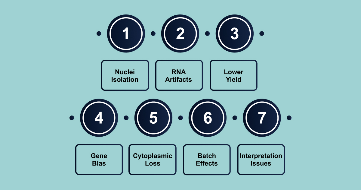
Single-nucleus RNA-seq (snRNA-seq) enables access to fragile or archived tissues but introduces technical challenges distinct from whole-cell methods:
- Intact, Contamination-Free Nuclei: Isolating clean nuclei requires balancing mechanical force and detergent strength. Tissue-specific barriers (e.g., myelin in the brain, extracellular matrix in the kidney and heart) require customized buffers, density gradients, or FACS cleanup.
- Ambient RNA and Droplet Artifacts: Floating transcripts from ruptured cells can distort profiles, obscuring rare cell types. Ambient RNA contamination, both non-nuclear and nuclear, requires specialized algorithms (e.g., CellBender, SoupX) for mitigation, though performance varies.
- Lower Molecular Yield and Data Sparsity: Nuclei contain much less mRNA than whole cells, leading to fewer UMIs, higher drop-out rates, and poor precision for low-abundance genes. At least 500 nuclei per cell type and deeper sequencing are needed for reliable differential expression.
- Gene-Length Dependent Bias: Long genes with many introns (e.g., KCNMA1, AK5) are over-represented, while short genes (e.g., CRABP2, TM4SF1) are under-represented, distorting analysis. Length-aware normalization is recommended to correct this bias.
- Loss of Cytoplasmic Transcripts: Cytoplasmic transcripts (e.g., ribosomal protein mRNAs) are under-represented, particularly in markers like microglial activation (APOE, SPP1, CD74), limiting disease state detection, especially in tissues with active mitochondrial or stress responses.
- Batch Effects and Data Integration: Differences in read composition (e.g., high intronic fraction, fewer mitochondrial reads) cause strong batch effects. Joint snRNA-seq and scRNA-seq data integration requires batch correction models (e.g., Harmony, scVI) for accurate cell type classification.
- Interpretation Pitfalls: Ambient RNA can cause false “transition” clusters, especially in neurons, and low RNA content in immune cells (e.g., neutrophils, activated microglia) may result in missed subtypes. Mitochondrial and ribosomal RNA filters in scRNA-seq are unreliable for nuclei due to natural depletion in these classes.
snRNA-seq requires careful handling of ambient RNA, gene-length biases, and tissue-specific challenges for reliable data interpretation. However, advancements in RNA sequencing technologies, such as those provided by Biostate AI, help streamline these processes and make them more accessible.
How You Can Overcome the Challenges of RNA-Seq with Biostate AI?
Biostate AI offers a comprehensive RNA sequencing solution that handles every step, from sample collection to delivering actionable insights. Whether you’re working with fresh, frozen, or archived samples, our expertise in RNA sequencing ensures that your data is accurate and reliable.
Here’s what we offer:
- Unbeatable Pricing: High-quality RNA sequencing starting at just $80/sample.
- Rapid Turnaround: Receive results within 1-3 weeks.
- Complete Transcriptome Insights: Comprehensive RNA-Seq covering mRNA and non-coding RNA.
- AI-Driven Analysis: Our OmicsWeb AI platform provides powerful, intuitive insights without the need for coding expertise.
- Minimal Sample Requirements: Works with as little as 10µL blood, 10ng RNA, or 1 FFPE slide.
- Low RIN Compatibility: Compatible with RNA samples having a RIN as low as 2.
- Multi-omics Support: Handles RNA-Seq, WGS, methylation, and single-cell sequencing data.
By integrating both cutting-edge technology and an efficient, cost-effective approach, we provide the reliable, actionable data that researchers need to drive forward their discoveries.
Conclusion
scRNA-seq and snRNA-seq bring unique advantages and challenges. While scRNA-seq is ideal for analyzing fresh, dissociable tissues, snRNA-seq excels in handling more complex or archived samples, providing researchers with a deeper understanding of gene expression.
Ultimately, the choice between these technologies depends on the specific research goals and the types of samples being studied.
To navigate these challenges and make RNA sequencing more accessible, Biostate AI provides a comprehensive solution, offering everything from sample collection to insightful data analysis.
With our AI-driven platform and cost-effective pricing of $80/sample, we are designed to simplify RNA sequencing for researchers, ensuring precise, reliable, and actionable results.
If you’re looking to take your RNA sequencing to the next level, get in touch with us today to discuss your research needs and receive a personalized quote.
FAQs
1. How do bioinformatics pipelines address the unique challenges of snRNA-seq data, such as contamination from ambient RNA and low RNA content per nucleus?
snRNA-seq data can be particularly susceptible to contamination from ambient RNA released during nuclei isolation, which can distort biological signals. Advanced quality control tools, such as valiDrops, use adaptive thresholding and clustering-based approaches to identify and filter out low-quality nuclei and barcodes affected by contamination, thereby improving the fidelity of downstream analysis.
2. What are the implications of using nuclear versus cytoplasmic RNA for downstream analysis?
Since snRNA-seq captures primarily nuclear transcripts, some cytoplasmic mRNAs and post-transcriptional modifications may be underrepresented. This can influence the detection of certain gene expression signatures and may affect analysis that rely on full transcriptome coverage, such as alternative splicing or allelic expression studies.
3. How does the choice of nuclei isolation protocol impact the representation of cell states and subtypes in complex tissues, especially for archived or frozen samples?
Optimized nuclei isolation protocols are critical for preserving the diversity of cell types and states present in the original tissue. For example, protocols tailored for long-term frozen brain tumor tissues can enable more accurate identification of rare or fragile cell populations that might be lost with standard approaches.
4. In what ways are emerging computational tools integrating multi-omic data with snRNA-seq to provide a more comprehensive view of cellular identity and function?
The integration of snRNA-seq with other single-nucleus omics (such as ATAC-seq for chromatin accessibility) is an active area of research. These multi-modal analysis require sophisticated bioinformatics pipelines capable of aligning and interpreting disparate data types, enabling deeper insights into gene regulation, epigenetic states, and cellular heterogeneity